

Comprehensive analysis of macro-modern ultrasound: scope of screening and clinical significance
Introduction: recognizing the importance of large-scale terraform screening
In the middle of a pregnancy, each prospective parent will face a critical prenatal check-up — the loudspeaker. This examination, an important component of the modern prenatal diagnosis system, carries with it the expectations and concerns of countless families. In clinical practice, most pregnant women undergo this systematic screening during the 20-24 weeks of pregnancy, the core value of which is to provide professional assessment of foetal structural development。
It is worth noting that the professional name of this examination is "current obstetric ultrasound examination of level iii, which is also commonly referred to in the medical literature as "supersound examination of the foetal system" or "targeted prenatal ultrasound examination". What is commonly referred to as "extreme deformities" stems mainly from their extensive scope and vital clinical significance. This examination not only takes a long time (usually 30-60 minutes) but also covers the development of the various organs of the foetus and is an integral part of the prenatal screening system。
Technical principles and implementation points for large-scale deformities screening
At the technical level, the high-resolution colour doppler ultrasound technique is used for the screening of large row malformations. This advanced imaging method can penetrate the abdominal wall of the mother and clearly shows the anatomy structure of the intrauterine foetus. In the course of the examination, the ultrasound physician systematically scans and evaluates the organ systems of the foetus in accordance with standardized operating procedures。
The optimal window of time for the examination is set at 22-24 weeks of pregnancy, the selection of which is based on multiple scientific considerations. At this point, the organs of the foetus are essentially fully developed and of moderate size; the abundance of sheep water provides a good acoustic window for ultrasound imaging; and the incomplete calcification of the foetus's bones reduces the barrier to acoustic penetration. These factors together constitute the "gold window period" for structural screening。
In practice, examining physicians require professional qualifications and extensive experience. They must be skilled in the acquisition techniques of the various angles of the foetus and be able to accurately identify the differences between normal and unusual anatomy structures. Examination of the stability of the environment, the sophistication of the equipment and the degree of co-operation of pregnant women have important implications for the accuracy of the results。
Specific content and scope of screening for large-scale malformations
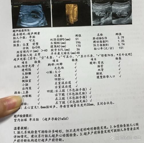
The system of content for mass deformations is comprehensive and covers almost all organs of the foetus. This examination is like a full "medical examination" of the foetus in the womb, each with its own specific assessment criteria and methods。
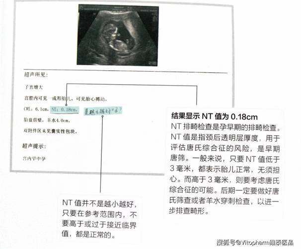
In terms of brain examination, doctors focus on the integrity of the skull radial ring, the structure of the mid-brain, the shape of the side brain, the position of the cortex, the development of the small brain, etc. These assessments can help to detect major nervous system anomalies such as brainlessness, severe cerebral fluids and deformities. It is worth noting that even experienced physicians may find it difficult to detect certain minor brain structure anomalies, which is one of the inherent limitations of the examination。
The evaluation of facial structure includes indicators such as eye symmetry, nasal bone development and upper lip continuity. These observations are important for the screening of abnormalities such as lasagna. It needs to be clear, however, that the assessment of facial microstructures, influenced by factors such as foetal heights, may be difficult, which is why some minor facial anomalies may have been omitted from the initial examination。
Cardiac examination, which is the focus of the large platoon malformations, typically includes the assessment of multiple standard tangents, such as the quartetal trajectories, and the left and right ventricular cleavages. These cuts allow doctors to screen for most serious congenital heart diseases, such as monocardials, large blood vessels, etc. It must be recognized, however, that there is still room for improvement in detection rates for certain complex heart malformations, especially those that do not involve significant structural changes。
Clinical value and detection capability for large-scale deformities
In terms of clinical value, the most significant advantage of large-scale malformations is the ability to detect structural malformations that are serious and could endanger the life of the foetus. Once discovered, these anomalies can provide an important basis for subsequent clinical decision-making, including whether further diagnostic examinations are required, whether termination of pregnancy is considered and whether special delivery plans are needed。
In the nervous system, the detection of major malformations, such as brain incompetence and severe cerebral water, is close to 100 per cent. These deformities tend to have typical ultrasound manifestations and have significant prognosis effects on the foetus. For neural tube defects such as spinal fractures, the sensitivity of the examination is also relatively high, especially in those cases where there is a marked continuity disruption of the spine。
The screening effects of cardiac malformations show greater variation. The overall detection rate for severe heart malformations is estimated at 60-80 per cent. This figure reflects the complexity of the heart examination, which is influenced both by the resolution of the ultrasound technology itself and by factors such as foetal position and timing. Some minor heart abnormalities, such as missing small room spacing, may be difficult to detect in the middle of pregnancy。
Abdominal defects, such as umbilical swelling and abdominal fractures, usually have a high detection rate in the major deformities examination due to their typical external performance. These anomalies are often accompanied by characteristic ultrasounds, such as an outburst of abdominal cavity, which enable doctors to make judgements relatively easily。
Analysis of the limitations of the large platoon malformations
Despite the important clinical value of large-scale malformations, we must be conscious of its inherent limitations. These limitations derive mainly from technical principles, fetal development characteristics and timing。
The most notable limitation is that the examination is essentially a morphological assessment and cannot detect functional anomalies. This means that even if all organ structures of the foetus are shown to be normal, the possibility of visual, hearing or neurological impairment after birth cannot be completely ruled out. These functional problems often require specialized post-natal examinations to be identified。
Chromosomal anomalies are another important screening blind zone. Although certain chromosome diseases may be accompanied by specific structural anomalies (e. G., possible heart defects in children with down syndrome), ultrasound tests themselves do not directly observe chromosomes. The diagnosis of chromosome abnormalities must rely on interventional prenatal diagnostic techniques such as water piercing。
The detection of microstructure malformations is also challenging. Problems with small structures, such as abnormality of the finger/toe, slight urea cracks, can be easily missed during mid-pregnancy ultrasound. These anomalies often require higher resolution equipment or more specialized inspection techniques to detect them。
There are also significant limitations in screening for late-mortality malformations. Certain diseases, such as some types of cerebral water, digestive barrier, may occur in late pregnancy or even after birth. This means that even if the results of the major malformations are normal, the possibility of subsequent development problems cannot be completely ruled out。
Professional interpretation and clinical response to inspection reports
Professional medical interpretation is essential when reports of major malformations are available. The professional terms and descriptions in the report require a comprehensive analysis by experienced obstetricians to arrive at an accurate clinical judgement。
When descriptions such as "xx structure is not clear" appear in the report, it is usually suggested that satisfactory observations could not be obtained for technical reasons. This does not imply an anomaly, but may require a review to obtain more complete information. The reference to "recommended review/follow-up" tends to suggest the need for dynamic observation of certain suspicious manifestations, which are common clinical prudence。
For specific anomalies mentioned in the report, such as the clearly diagnosed "literal crack" or "vertebrae", pregnant women and their families should communicate with obstetricians in a timely manner. Depending on the exceptional circumstances, the physician recommends further examination options, with a detailed explanation of possible pre- and treatment options。
In particular, it is important to emphasize that in no case is it recommended that non-professionals interpret ultrasound reports themselves. The fragmentation and unprofessional nature of network information can easily lead to misunderstanding and excessive anxiety. Professional medical advice has always been the most reliable source of information。
Distinguished from other obstetric ultrasound tests

Each level of ultrasound has its own specific clinical orientation and value in the complete system of prenatal examinations. Understanding these differences will help pregnant women to organize their screening programmes rationally and avoid unnecessary duplication or omissions。
Level i ultrasound, as the most basic examination, is used primarily to confirm pregnancy status, assess foetal survival, measure basic growth parameters, etc. Such inspections usually do not involve detailed structural assessments, are relatively simple and can be performed during routine production tests。
Level ii ultrasounds already have some structural screening capacity to observe the development of the principal organs of the foetus. In many medical institutions, this level of examination already allows screening for most of the obvious structural anomalies. The content and level of detail of the examination is between levels i and iii and forms an important part of the antenatal screening system。
Level iii ultrasound, which is the focus of this paper, represents the highest level of prenatal structure assessment. Such inspections are usually conducted by experienced experts, using high-resolution equipment, with a high standard of content and depth. This applies in particular to high-risk pregnancies or suspected foetal abnormalities。
Clinical advice and summary
On the basis of the above analysis, we make the following professional recommendations for the examination of macro-tetanus:
First, it is recommended that all pregnant women complete this examination during the 20-24 weeks of pregnancy. The selection of this window has been fully clinically validated, balancing the accuracy of the examination and the timeliness of clinical intervention. Early or late inspections may affect the reliability of the results。
Secondly, the results of inspections should be viewed in a rational manner. Attention must be paid both to any unusual signs detected by the examination and to the understanding that normal results do not guarantee the absolute health of the foetus. This balanced mentality helps to reduce unnecessary anxiety while maintaining the necessary vigilance。
Finally, we stress the importance of professional medical guidance. Both pre-inspection preparation, co-operation during the examination and post-inspection consultations should be conducted under the guidance of a professional physician. Medical institutions should also provide pregnant women with adequate counselling services to help them understand the meaning and results of examinations。
The value of mass deformities as an important component of modern antenatal care has been widely recognized. Through scientific awareness and rational application, this technology will bring health and security to more families。