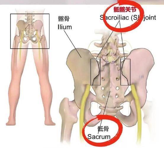
The gillbone, located on both sides of the human pelvis, is one of the important bones that form the pelvis, and acts primarily to support the body and to protect the pelvis internal organs。
1. Anatomy location
The gillbone occupies a prominent position in the human skeletal system, specifically in the area below the waist that connects to the top of the base of the thigh. It is the largest of the three bones that make up the pelvis, and the other two are sciatic and shameful. When you touch your body, the most prominent bone on either side of your waist is referred to as a “skull axle” or the upper part of your waist, which is referred to in medical terms as gill. This part, while standing or walking, has the key task of focusing on the transfer of the upper half of the body to the lower limb。
2. Form structure
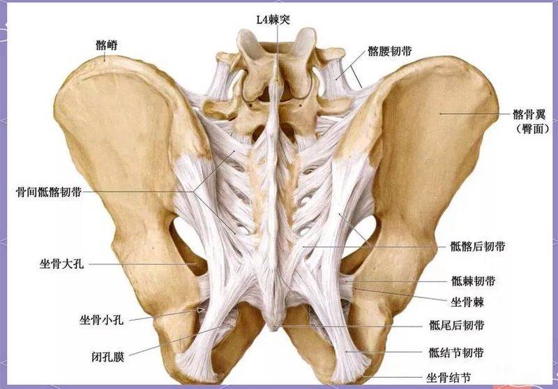
In morphological terms, the osteoporosis presents an irregular sector or wing structure divided into thick osteoporosis and flat osteoporosis. The osteoporosis is involved in the upper part of the body, which forms a hip joint with the bone and ensures the flexibility of the movement of the lower limb. The osteoporosis wing spreads upwards, with the internal cavity dent forming a cavity, which is a point attached to the muscular muscle; it is condensed outside for the hips. This unique structural design both ensures the pelvis's robustness and provides a wide area of attachment to powerful muscles to sustain the human body's straight posture and motion function。
3. Physical function
The core function of the ostrich is to build a solid pelvic ring that effectively protects vital organs such as bladders, rectums and female uterus and ovaries in the pelvis from external force damage. At the same time, as a mechanical bridge between the torso and the lower limbs, the ostrich is responsible for spreading the gravity from the spine evenly to the lower limbs on both sides and for maintaining the stability of the centre of gravity. In addition, the hemorrhoid marrow is rich in the gillbone, which is the main place of blood creation in childhood and adolescence and, although partially transformed into yellow marrow in adulthood, retains a certain potential for blood production in the light of specific physiological needs。
4. Common pathologies
The osteoporosis area may suffer from discomfort or disease for a number of reasons, including cystosis, most common in the case of women with dysentery, in the form of pain in the waist; second, fractures of the osteoporosis, usually caused by high-energy traumas such as car crashes or falling in high places, accompanied by severe pain and restriction of activity; and second, tumour pathologies, which may be caused by cystomas or cancer cells transferred from other parts, which may violate the osmosis, causing local swelling and persistent pain. These rational changes often require diagnosis through video tests and timely and targeted treatment。
5. Access methodology
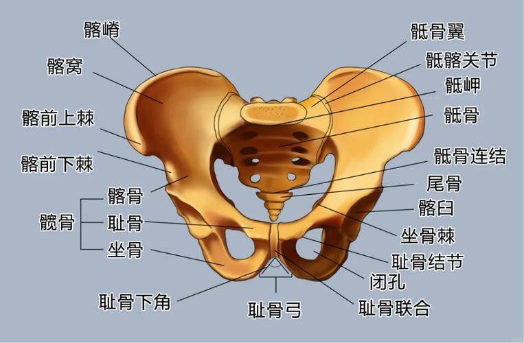
Ordinary people can sense the position of the bone through simple self-disturbation. Take a stand, hand down naturally, and put your hands on the most prominent bone markings on both sides of the waist, slide along the bone down, and touch a long arc ridge, which is the arc ridge. Touches on both sides of the abdomen along the gills, touching the thorns in front of the gills, which are the attachments to many muscles and lurches and are important signs of clinical leg length and positioning. In this way, the approximate distribution of the gillbones in the body table can be seen in a visual way。
In daily life, maintaining the right positions and positions helps to reduce the burden on the pelvis and pelvis areas and to avoid bad habits such as long-term single-side weighting or gill legs. Moderate core muscle exercise, such as tablet support or bridge exercise, enhances the muscle strength and pelvis stability around them. In the event of continued pain, swelling or movement disorders in the osteobrane area that are not detected for unknown reasons, do not carry out self-massing or heat dressing, visit the hospital in a timely manner to identify the cause of the disease through a professional examination, so as not to delay the situation affecting normal life。