


The path of scientific research begins with observation that, in the exploration of the microworld, electron microscopes and optical microscopes, like the extension of our eyes, lead us into a very different tiny universe。
The microworld has always been a fascinated mystery, and humankind has longed to find out since ancient times. From the invention of optical microscopes in the 17th century to the advent of electron microscopes in the 20th century, our ability to observe the microworld continues to grow。
While both types of microscopes seek to reveal microstructures that are invisible to the naked eye, they differ in their rationale, performance and application. The digital microscope, as a product of modern micro-technology development, further expands the application boundary of micro-observation by combining the ease of optical microscopes with the digital advantage of electronic microscopes. For scientists and engineers, understanding their differences and making the right choices is the first step in exploring the microworld。
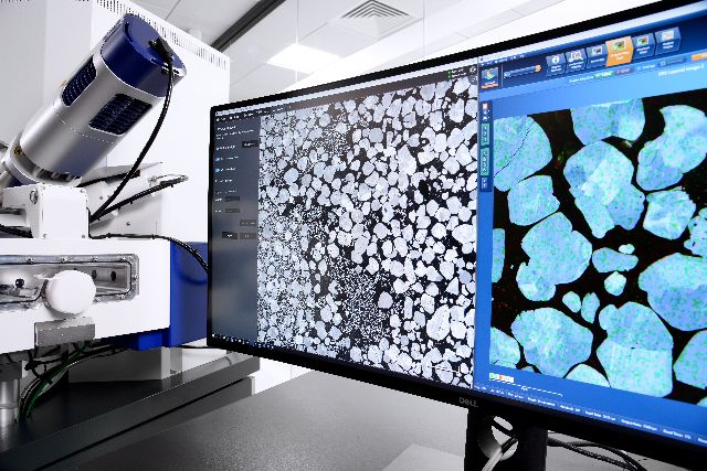
I. Rationale, nature of electronics and light
Optical microscopes are the first type of microscopes that most people have been exposed to during their student years and are based on a combination of visible light and glass lenses. Optical microscopes use light across a series of lenses to magnify small objects, usually consisting of components such as eyeglasses, mirrors, microscopes and light sources。
The light source usually comes from visible light bands with wavelengths in the range of 400-700 nm, which also determines the theoretical maximum resolution of optical microscopes of approximately 0. 3 micrometres。
The process of imaging of optical microscopes begins with light coming out of the light, which focuses on the samples through the lens, and is then captured and formed into primary amplification through part of the light through the sample, which is then further amplified through the lenses and observed by our eyes。
Unlike optical microscopes, the revolutionary nature of electron microscopes is that they use electronic beams rather than visible light as a “light source”. According to the material wave theory proposed by debrovage, electronics are volatile and high-speed electronic wavelengths are much longer than visible light waves。
Electronic microscopes use electromagnetic lenses rather than glass lenses to control and focus electronic beams. These electromagnetic lenses deflect and focus the electron path through a precision-controlled magnetic field to form magnified images。
The electron microscopes are divided into two main categories: a penetrating electron microscope and a scanning electron microscope。
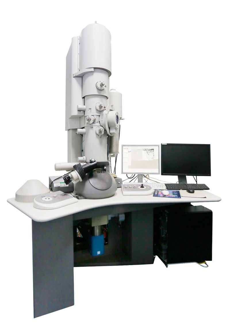
Transmitting electron microscope (tem)
The mode of work of the transient electron microscope (tem) is similar to that of the optical microscope, which allows the beam to penetrate the sample before imaging。
The tem, however, needs to accelerate the electron to near light speed, and the electron collision with the atoms in the sample produces a stereo-angle dispersion, the size of which is associated with the density and thickness of the sample, resulting in a dark and dark image。
Tem is able to provide information on the internal structure of the sample, with resolution up to atomic level, but requires that the sample be very thin, usually not exceeding 100 nm。
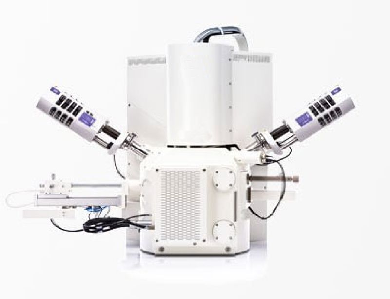
Scan electronic microscope (sem)
The scanning of the electron microscope (sem) uses completely different working principles. It constructs the image through a point-by-point scanning of the sample surface by focused electronic beam and then by detecting secondary electronic or back-scattered electronic signals inspired by the sample surface。
The electronic beam produced by the sem electronic gun is focused on very fine probes by the electromagnetic lens system, which, under the control of the scanning ring, are scanned on the surface of the sample at a certain time and in sequence。
This mode of imaging enables sem to obtain three-dimensional stereoinformatic information on the surface shape of the sample, which, although usually lower than tem, has a deeper and more intuitive 3d visual effect。
The digital microscope, as an emerging member of the family of microscopes, adopts a different design concept from the traditional microscopes. It brings together fine optical microscopes, advanced photovoltaics and liquid crystal screens。
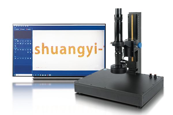
Digital microscope
Digital microscopes convert physical images that are seen in microscopes through numerical simulations, making them visible on screens or computers with microscopes. This design leaves the digital microscope without a goggles, and can be used to export real-time images to monitors through cameras and amplified optical devices, enabling multiple simultaneous observation of images。
Ii. Resolution and amplification capabilities, across quantitative scales
Resolution is one of the key indicators of microscope performance, which represents the minimum distance that the microscope can distinguish between two adjacent points. In this core parameter, the electron microscope shows an overwhelming advantage。
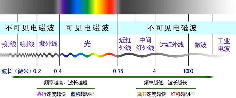
Optical microscopes are usually not capable of exceeding 0. 3 μm in resolution due to the limitations of visible wavelengths. This means that if the distance between two objects is less than 0. 3 μm, they will be blurred under the optical microscope and cannot be distinguished from two separate entities。
The resolution of the transient electron microscope was as high as 0. 1-0. 2 nm, which was thousands of times higher than the optical microscope. Such resolution is sufficient to allow us to observe the structure of just one atom column and to reveal the most fundamental composition of the substance。
In terms of amplification, ordinary optical microscopes can typically be magnified up to a maximum of 1,000 times, up to a maximum of 1,600 times by optimizing optical components。
The magnification of transient electron microscopes can be tens of thousands to millions of times, and can easily reveal micro-areas beyond the reach of optical microscopes。
The magnification factor for the digital microscope is calculated differently from the traditional optical microscope. For digital microscopes, because the image is observed on screen, the magnification factor is the multiplier of the lens optical magnification factor and the display size。
The total magnification factor for the digital microscope can be calculated using the following formula, which is equal to the monitor magnification factor multiplied by the optical magnification factor. Some digital microscopes provide 20 to 7,000 times the optical zoom amplification range, which is a high magnification factor that enables them to respond to the needs for microanalysis observed from macros。
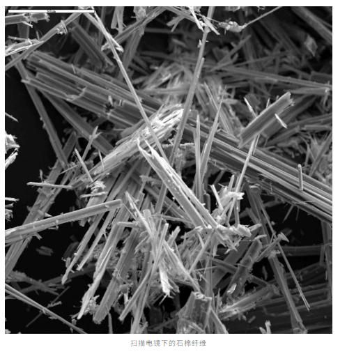
The underlying cause of this huge gap lies in wavelength differences. The optical microscope uses visible wavelengths of 400-700 nm, while the electronic beams used in the electron microscope are much shorter after acceleration。
The wavelength of the electron is associated with the acceleration voltage according to the deburo formula, and the higher the acceleration voltage, the shorter the electron wavelength. The nature of this wavelength allows the electron microscope to break through the distillation limits of the optical microscope and open the door to the nanoworld。
Iii. Technical options between sample preparation and processing and simplicity
There are also significant differences in sample preparation requirements between microscopes, which are directly related to their applicability and efficiency in actual research。
Sample preparation for optical microscopes is relatively simple. For common biological samples, it is usually only required to be fixed on the tablet, sometimes dyed to increase the contrast。
Live-cell observations are simpler and can be observed directly in the field or under the differential microscope, without complexity. This simple preparation process makes optical microscopes particularly suitable for teaching and rapid testing。

In contrast, the electronic microscope sample preparation is complex and time-consuming. Transradioscopes require that samples be very thin and usually require the preparation of ultra-scaling slices about 50 nm。
The production of these ultra-slice slices requires the completion of the super-slice-slice machine, which is extremely sophisticated. Biological samples are also subject to a series of treatments, such as double fixation of peptide and acrylic acid, resin packaging, etc。
For sample scanning of mirrors, there is a need to go through a fixed, dehydrated, critical point drying and, finally, to spray thin sheeted membranes on the sample surface to increase the secondary electronic launch。
These complex preparation processes require professionals to operate and, more importantly, make it impossible to observe samples of living organisms. The complexity of sample processing directly determines the applicability of the two microscopes in different scenarios。
Digital microscopes have a unique advantage in the preparation of samples, which can be observed without the need to dismantle and process the target. For users in the field of industrial testing, this feature greatly simplifys the sample preparation process and does not require complex pre-sampling, as is the case with electronic microscopes。
Iv. Applications, each having its own area of application
Although electron microscopes have an absolute advantage in resolution and magnification, they have not been eliminated as a result, and they play an irreplaceable role in different applications。
Optical microscopes play a central role in the life sciences by virtue of their non-intrusive character. It can track dynamic processes within active cells in real time, such as observation of morphological changes in neurotic synapses in neuroscience, and cytological splits in developmental biology that track zebra fish embryos。
In medical diagnosis, hospital pathology relies on optical microscopy analysis tissues to recognize cancer cell forms through high-resolution imaging and assist with tumour spectrometry and stratification。
In addition, optical microscopes are widely used because of their simplicity and relatively low cost in material science, geology and education。
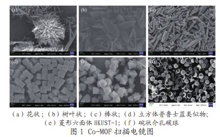
Electronic microscopes play a key role mainly in nanoscale research. In the field of material science, it is used to study the internal microstructure of materials, analyse particle sizes, phase composition, growth orientation, crystals and crystal defects, etc。
In the semiconductor industry, transient electron microscopes are used to analyse faults and mixing distributions in semiconductor materials and to assist in the development of next-generation chips。
Biostructural studies also involve electron microscopes, particularly refrigerated mirrors, which, by cooling samples to liquid nitrogen temperatures, can observe temperature-sensitive samples such as proteins, biochips, etc., significantly reduce the damage of electronic beams to samples。
By virtue of its unique advantages, the digital microscope has found its own application location in many areas. In the field of industrial detection, digital microscopes are widely used in the electronic manufacturing industry for the detection of integrated circuits, semiconductors, smt and pcb circuit boards。
Precision parts defects, cracks and data measurement analysis are also used in the precision machinery industry, and print quality testing and ink observation analysis are used in the printing industry. In the textile industry, quality control is used。
V. Summary, complementary micro-exploration tools
Electronic microscopes and optical microscopes are two important tools for human exploration of the microworld, each of which plays a unique role at different scales。
The electro-microscope, with its super-high resolution, allows us to see the mysteries of the atomic world, contributing to breakthroughs in material science, nanotechnology and structural biology。
Optical microscopes continue to play an irreplaceable role in life sciences, medical diagnosis and education through their non-intrusive, live observation capabilities。
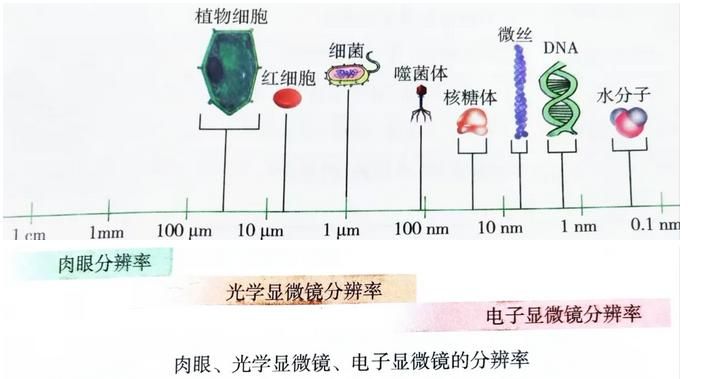
Technological advances are blurring the boundaries of the two microscopes. The breakthrough in ultra-resolution fluorescent microscope technology allows optical microscopes to break the distillation limit into nanoscale observation。
Refrigeration mirrors, on the other hand, allow electron microscopes to observe biological samples closer to their physiological state。
Electronic microscopes and optical microscopes are not competitive but complementary instruments of exploration in the march of scientific research. Understanding their differences and advantages and making informed choices based on specific research needs will help us move further along the path of exploring the microworld。
Whether it reveals the mystery of life or promotes the development of material, both types of microscopes will continue to serve as the eyes of science, leading us into the invisible wonders of the world。