If you want to know what an ekg looks like, first you have to know what an ekg is。
What's an ekg
An electrocardiogram (ecg or ekg) is a technique for recording from the body chart, for each heart cycle, the graphs of electrical activity. During each heart cycle, the pace point, the heart room, the heart room is excavated, accompanied by changes in bioelectricity, and many forms of electrostatic changes are elicited from the body chart by electrocardiographers。
The principles of ekg generation
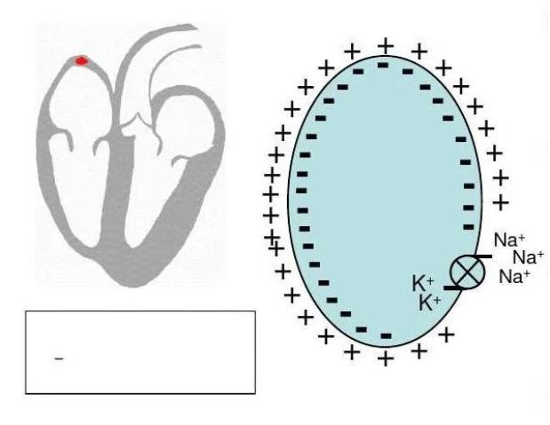
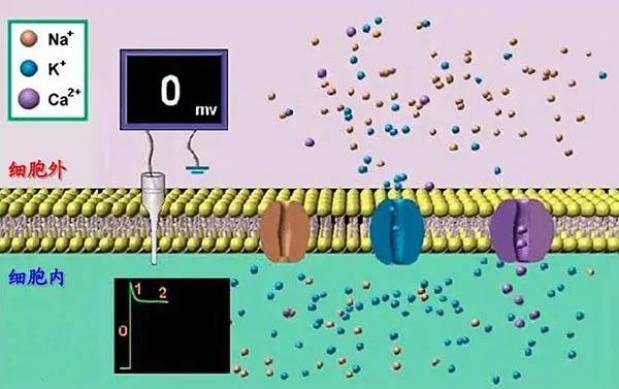
Here's how the electrocardiogram works. Acoustic medicine has been found to be a semi-permeable membrane in the heart, statistically organized outside the membrane with a certain number of positive charged anion, within the membranes with the same number of negative charged anion, and above the membrane level, called polarization. In the static state, because of the polarization of all the ocular cells in the heart, there is no difference in the level of the electron recorder's image of the electrocution curve, i. E. The level line of the ekg. When the myocardial cell is irritated with a certain intensity, the membrane permeability changes and a large number of anion enters the membranes in a short period of time, resulting in a negative correction of the inner membrane electrical level, a process known as depolarization. For the heart as a whole, changes in the level of electrons during the process of depolarization of myocardial cells from the inner membrane to the outer membrane sequence are described by the electrostatic curve as the depolar wave, i. E. The p-wave and the qrs wave of the heart chamber on the ekg. After the completion of the diaphragm, the cell membrane excretes a large number of anion, which causes the inner membrane level to revert from a positive to a polarized state, which is carried out from the outer membrane to the inner membrane, known as the repolarity. The changes in the level during the repolaration of myocardial cells are captured by the current recorder, known as the contour wave. Due to the relative slowness of the repolarity process, the repolarity is lower than the detoxification wave. The cylindrical electrons of the heart chamber are not easily identifiable when they are very low-waved and buried in the cleavage wave of the heart chamber. The contours of the heart chamber are shown in t-waves on the chart of the body. Once the whole of myocardial cells are re-introduced, the polarized state is restored again, there are no power levels between the various myocardial cells, and the electrostatic chart of the body is recorded as the equivalent. We'll see what the ekg looks like through an electrocardiogram。
Method of recording electrocardiograms
Ekg
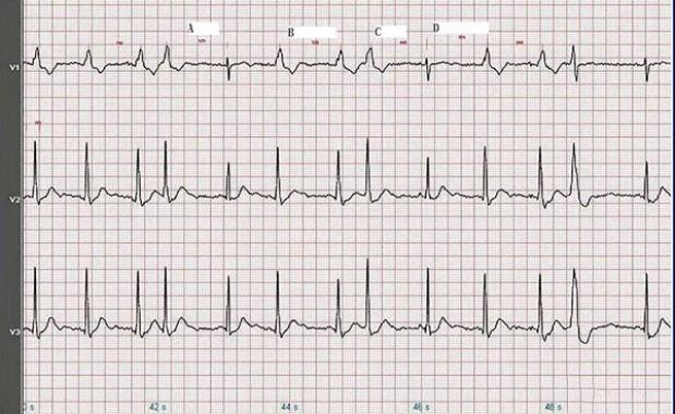
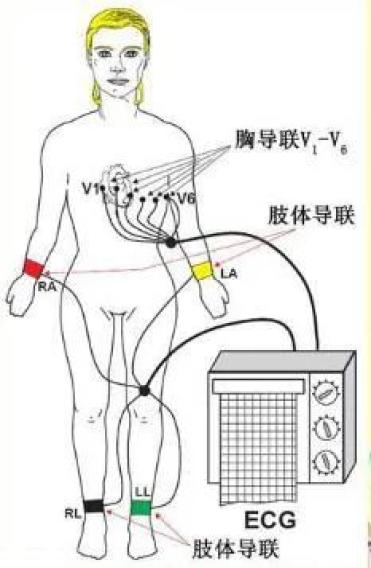
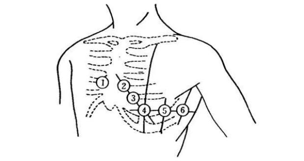
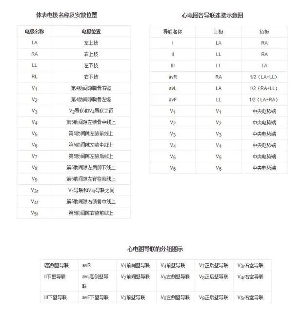
Acoustic medicine has learned that when the heart is constricted and constricted, there is a small bioelectricity that can be detected and recorded from different parts of the body surface using electrocardiograms. The electrodes are placed in different parts of the human body in the image of the electrocardiogram, which is used to record and react to the electrical activity of the heart. The location of the heart electrode is shown below. Only four body conductor electrodes and v1-v66 chest pre-conductor electrodes are normally installed during routine electrocardiograms, recording regular 12 conductor electrodes。
A different conductor is formed between the two electrodes or between the electrodes and the central power tip to record the electrical activity of the heart through a direct link to the positive-negative current meter. The two electrodes form a bipolar steering link, one positive and one negative. The bipolar body conductors include i, ii and iii; a unipolar conductivity is formed between the electrodes and the central power tip, at which point the detection power is extremely positive and the central power edge is negative. Avr, avl, avf, v1, v2, v3, v4, v5 and v6 are unipolar. Since avr, avl, avf are far from the heart and the difference in the level of power is too small when the central end is negative, the sum of the bits of the two limbs other than the probe of the electrodes is applied. This record adds avr, avl, and avf fuses, which are also referred to as unipolar cavities。
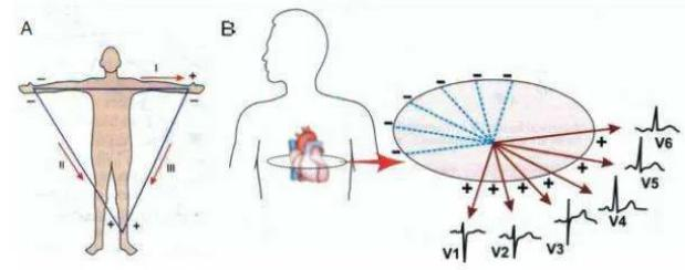
The body conductor system reflects the heart level projection on the vector side. Including i, ii, iii, vr, vl and vf. The chest frontal conductor system reflects the heart level projection including v1, v2, v3, v4, v5, v6. They are further grouped to reflect electrical activity in different parts of the heart。

Central power: also known as the wilson central power end, it is created by connecting ra, la, ll electrodes through a resistance network, representing the average voltage of the body. This voltage is close to a great value (i. E. Zero)。
Cardiograph log paper
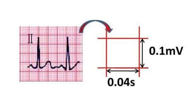
The ekg records a curve of voltage over time. The electrocardiogram is recorded on the coordinate paper, which consists of a small grid of 1 mm wide and 1 mm high. Cross-coordinates indicate time and vertical coordinates indicate voltage. It is usually recorded at 25 mm/s paper speed, with 1 grid = 1 mm = 0. 04 seconds. (b) transcript = 1 mm = 0. 1 mv。
Composition of ekg
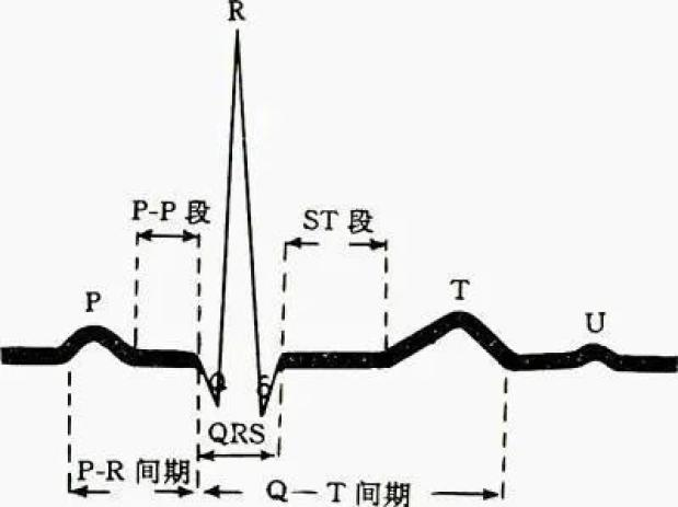
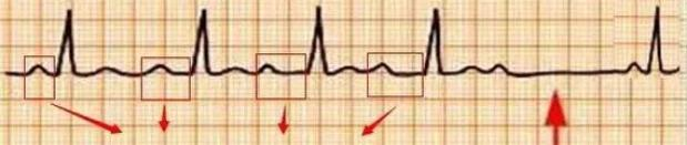
1. P wave
The normal heart's electro-excited starts at the end of the inn. Since the cylindrical knots are at the intersection of the right-heart and upper cavity veins, the agitation of the cylindrical knots is first transmitted to the right-heart, and via the room beams to the left-heart chamber, forming the p-waves on the ekg. Ppo represents the excitement of the heart, the excitement of the first half of the body, the excitement of the left half. The p wave time limit is 0. 12 seconds and the altitude is 0. 25 mv. When the room is expanded and the transmission of the two rooms is abnormal, the p wave can be shown as a high point or double-peaker。
2. Inter-pr period
The excitement is transmitted along the middle and back beams to the room. Owing to the slowness of the room's conductor, a ppr section, also known as the inter-pr period, was formed on the ekg. The normal pr period is 0. 12 to 0. 20 seconds. Watch out for delays in transmission to the heart room, as evidenced by the extension of the ppr period or the disappearance of the heart chamber after the p wave。
3. Qrs waves
Qrs waves are formed in the heartrooms of each of the two groups. The qrs band represents the heart chamber decomposition and has an exciting duration of less than 0. 11 seconds. Qrs bands are enlarged, deformed and time-limited when there is a conductive retardation, expansion of the heart chamber or thickness of the right-and-right branches of the heart。
Point 4. J
End of qrs wave, nodal point for start of st segment. On behalf of the total decompression of myocardial muscle cells。
5. St
All myocardial muscles have been detached for a period of time that has not yet started. At this point, the cardiac muscles of all sections are depolarized, and there is no electrical difference between cells. The st segment should therefore normally be on the level line. When a certain part of the myocardial muscle showed signs of ischaemic or necrosis, the heart chamber continued to suffer from electroplasm after the decomposition, as shown by the deviation of the st section on the ekg。
6. T wave
Then the t-wave represents the end of the heart. In the qrs main wave direction, the t-wave shall be the same as the qrs main wave direction. Changes in t-waves on the ekg are influenced by a number of factors. For example, myocardial insufficiency can be seen in t-wave low-level reverses. T-wave heights can be seen in high blood potassium, acute myocardial infarction periods, etc。
7. U wave
The u-waves, which are visible after the t-waves, are currently considered to be related to the re-establishment of the heart chamber。
8. Inter-qt period
It represents the time of the heart room from the cleavage to the restoration. The normal qt period is 0. 44 seconds. The concept of the corrective qt period (qtc) was introduced because the qt period was affected by the heart rate. One method of calculation is qtc=qt/√rr. The extension of the qt period is often associated with the occurrence of malignant cardiac disorders。
What about the ekg
The following is a detailed account of the electrocardiogram:
(i) value
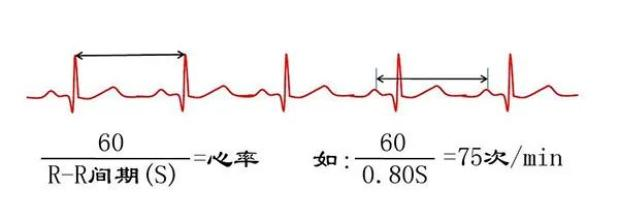
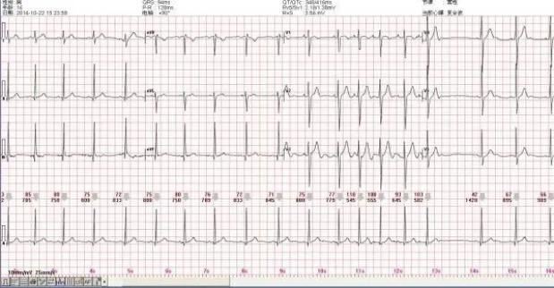
1 heart rate: normal 60-100。
Less than 60bpm's newspaper, “the momentum heart is too slow”, and more than 100 newspaper, “the momentary heart is too fast”。
2 p wave: 120 ms at normal time and double-peaker, reporting “diploon p wave” (sometimes visible, not much)
3. Pr breaks: normal time 120-200 ms。
>200ms newspaper, “at-time room transmission block”;
Qrs inter-period
5 rv5 > 2. 5 mv and/or rv5 + sv1 > 4. 0 mv (men)/ > 3. 5 mv (women), reporting “left room high voltage”, is very common! It is the left room strong performance, follow-up of medical history, high blood pressure, visual electrocardiograms, and the qrs band is significantly elevated。
6 p/qrs/t axis: xx/xx/xx, mid-30. -90. Report, “current axis left” (very common), 90. - 180. The "right side of the axial axis" (not much, but i am) is more specific, moderate, usually medium, and medium and above!
These values are shown on the ekg, so learn how the ekg looks, and understand the normal range of the above data。
Ii) see if there's an early fight
The qrs form, according to the medical knowledge of boeing, has a wide-ranging malformation called “room time” or “room time”, which is not properly identified with the sinus heart, and which is divided by 2 by the two normal waves of the rr period before and after it is equal to a normal rr period (which is difficult to understand, please overcome), with a round scale, which is not uniform. Reports are prepared on a regular or occasional basis, mostly as a “frequent and early twos/threes” and less than three in a minute。
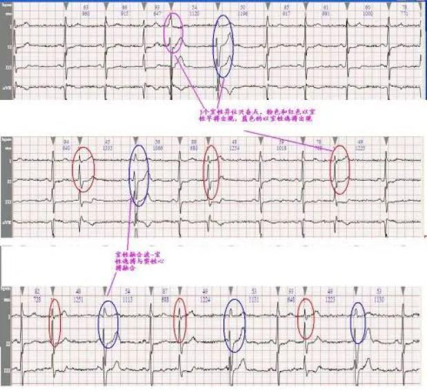
In addition, if there is an early pace, a fraction should be added to see how many of the fractions are, and if there is an occasional one, but not one, a dynamic electrocardiogram should be considered。
(iii) poor heart rate
If you can find a time difference of more than 120 ms in the rr period, it's not like you're going to fight early and you're going to get a "sympathetic heart disorder" that many people have. It is common for young and young people to be concerned with breathing, and it is mostly with breathing and not enough to be able to disappear. Patients often ask what this is about, and if the electrocardiograms are normal, except, they usually have no clinical significance, and sometimes my telegraphs。
(iv) see if there's room tremors, room pounces
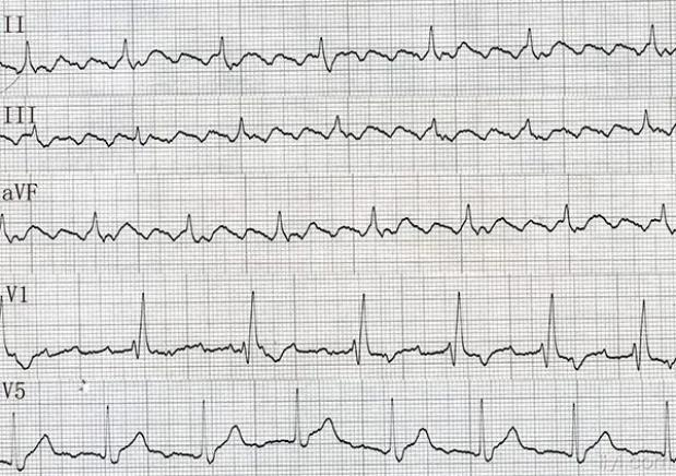
Theoretically, room tremors are normal p-waves disappearing, replacing them with different sizes and different forms of tremors, which are visible in constant v1. The normal identification of both can be seen as an absolute tremor. House tremors are more common than house puffs。
(v) right beam transmission retardation
If the qrss of v1 and v2 are in m or rsr ' form, the right beam transmission is blocked, with the qrs time limit >120 reporting a “full right beam beam beam beam beam beam beam beam beam beam beam beam beam beam beam beam beam beam beam beam beam beam beam beam beam beam beam >120 reporting a “full right beam beam beam beam beam beam beam beam beam beam beam beam beam beam beam beam” or otherwise, the reporting is incomplete。
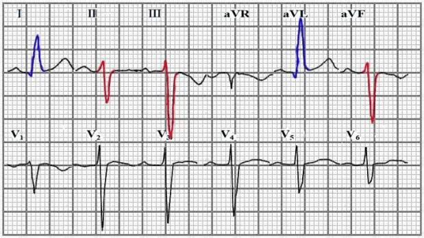
(vi) blockage of forward left branch transmission
Qrs of i, ii, avf is rs-shaped, on the left side of the axle, sometimes。
(vii) st, t-wave changes
At least one quarter of the patients examined reported “t-wave changes”, mainly in the form of low or upside down. In general, it is considered that: i, ii, v5 and v6 should be "t-wave reverse" if the t-wave amplitude is "t-wave" lower than 1/10 of the co-conductor r-wave。
With regard to st, it should be understood that st refers to the line between the endpoint of the qrs band and the beginning of the t-wave。
The position of the two cells following point j (start of the st segment) is defined as the "st section low" if only 0. 5 mv is moved down in any connection; and the "st segment elevated" is "0. 3 mv and/or v3>0. 5 mv and/or v4-v6> 0. 1 mv" on v1-v2。
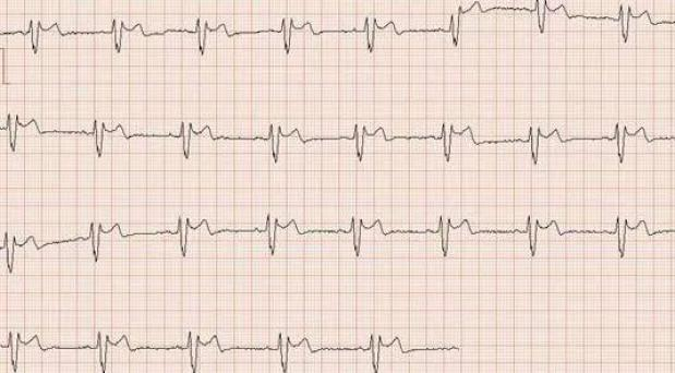
(ix) second degree i room transmission block
The p-waves always appear, but the longer the p-pr period, the longer a p-wave is, the more qrs is missing, the more qrs is circular. Wen's phenomenon. Pretty common。