

In recent days, professor y. Shrike zhang's team at harvard medical school has published an overview paper (inline) in the small magazine summarizing and looking forward to research developments in 3d bioprinting, including, inter alia, history and recent developments in bioprinting; progress in research into functional bioink to obtain the best biomimication structure; future directions for bioprinting; and potential applications of multiple bioprinters and their printing organizations currently available on the market。
The preface describes the inevitability and advantages of 3d cell printing. In the area of organizational engineering, there are many traditional ways of forming functional stereoorganizations, including three-dimensional support frames, self-equipment microengineering techniques and non-spare cell membranes. These methods have advantages but are not renewable in terms of high accuracy and control, and the 3d bioprinting technology developed in recent years can solve this challenge with unprecedented accuracy and precision by printing biological materials and cells into renewable stereostructures through intelligent programming equipment. Bioprinting techniques, bio-ink design and cell sources, in turn, determine whether the required biological structure can be successfully printed。
The first part of the text summarizes the working principles, strengths and weaknesses of photo fixation printing, ink-jet bioprinting, laser-assisted bioprinting, crowding out bioprinting, and electrostatic silk bioprinting。
Part ii summarizes the functional ink design for bioprinting, its mechanics, biological properties and electrical properties. Bioink underpins the engineering tissue and organ structure of 3d printing and is one of the most critical factors determining success in printing. Biological ink currently under development is alginate, fibrinogen, gelatin, collagen, chitosan, agarose, proronics, hyaluroNic acid (ha), gelma, peg and decelularized ecm (decm) and its mixed materials。
Part iii summarizes the origin of biologically printed cells. The membrane-filled stem cells (mscs) are more widely used in bioprinting, and fat stem cells (ascs), sheep stem cells (afscs), cow artery inners (baecs), embryonic stem cells (escs) and fully divided cells are also suitable for 3d bioprinting applications。
The fourth part discusses in detail recent research progress in bioprinting, including any complex form of biomimicry (figure 1), biomimicry (figure 2) with gradients and multimaterial structures, high-resolution bioprinting, in situ bioprinting (figure 3) and 4d bioprinting。
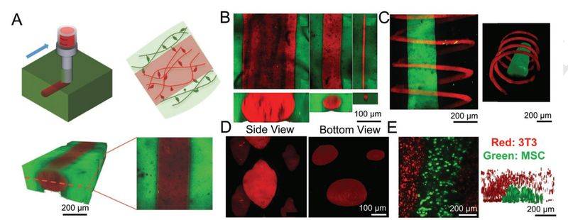
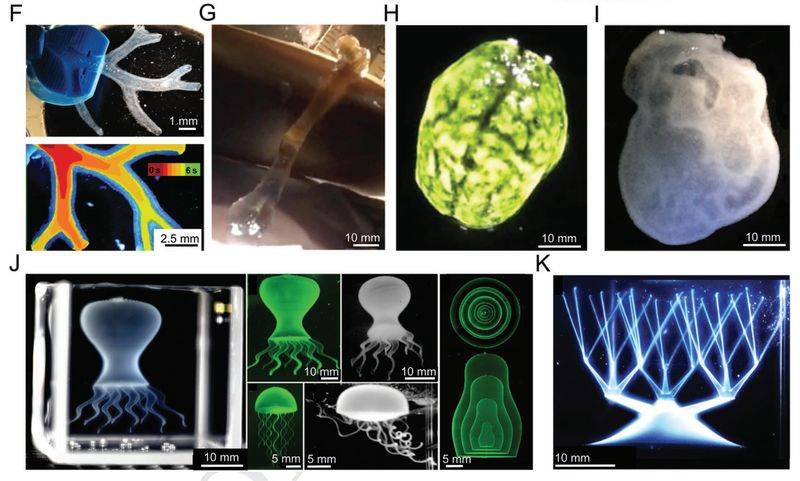
Figure 1 any form of biological printing in different shapes and structures
A) the rationale for injecting supermolecular ha-ad into the ha-ch matrix figure
B) pumping of fluorescent-labelled water gels with different needles medium
C) bioink ink threads for fluorescent tags, bioink spirals for rodanmin tags into unlabelled co-focal maps in hydrogels
D) co-focal maps of discrete elements in unmarked water gel injected incoherent ink from rodanmin markers
E) printing of inter-filled stem cell (green) focal maps in bioink supported by nih/3t3 cell fibre cell (red) hydrogel
F) models of the human right coronary artery and bioprinting structures based on a 3d magnetic resonance map of the embedding structure
G) models designed for bio-ink printing of human femurs and sodium algae to remove hydrogels
H) 3d bio-print brain structure
I) 3d bioprinting heart structure
J) model of 3d bioprinting octopus spectroscopy, multilayer structure
K) 3d bioprinting of a continuous network of hollow blood vessels
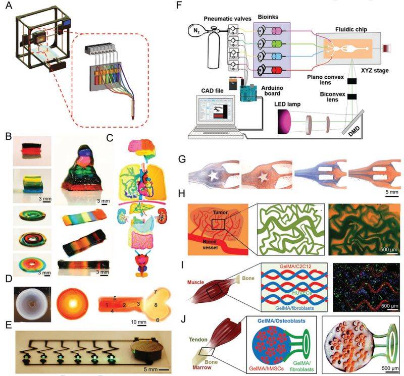
Figure 2 recent solutions for crowding out of multimaterials and cataract bioprinting
A) multimaterials consisting of 7 channels and a nozzle crowd out the biological printer rationale
B) biological printing structure of cubes, rings, pyramids, bar figure
C) bioprinting structures of different organs
D) bioprinting of gelma-hyroxyrophosphate structure for displaying bone induction gradients
E) bioprinted and connected circuit structure using gelma with different concentrations of carbon nanopipes (showed by the strength and weakness of green leds)
F) multi-material 3d plane with uv sources (385 nm), optical lenses and mirrors, dmd chips, microfluent devices figure
G) micro-meteoroid chip bio-ink injection interchange process in star-type, double-reconcil mode
H) motion (left), bio-print photomass, bio-printed tumour structure of gelma vascular tumours (right)
(i) momentum (left) of the skeletal muscle model, photomass (membracing) of the bioprint of the fluorescent micrograph of the gelma structure with c2c12 skeletal cells (red), fibre cells (blue) and vascular inner-skin cells (green)
J) momentum (left) of the skeletal embedding model, photomass (mode) of bioprinting, bright field optical of chromosomal structure of organisms with corresponding characteristics figure
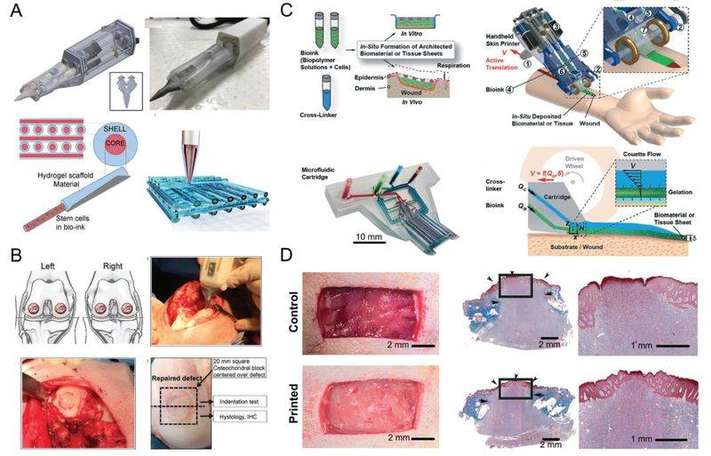
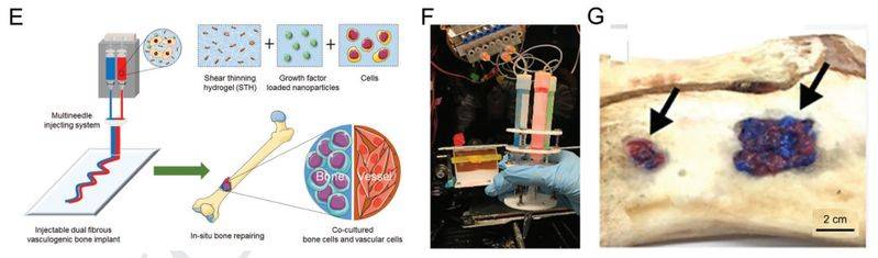
Figure 3 handheld in situ bioprinting solutions
A) the principle of printing pens for biology and photographs, and the principle of squeezing cells and ink at the bottom. Figure
B) treatment of full-layer cartilage defects with biological pens in the centre and side of the femur
C) momentum (top left), effect (top right), picture of bio-ink pens with different bio-ink combinations (low left) and their application to repair skin surface (low right)
D) summary of pictures of the control skin, the surface treatment group skin and the masson trichrome corroded tissues
E) the principle for in situ printing of ppl nanoparticles containing induction factors for the treatment of bone defects by encapsulating them with silicate cut-down water gels (sth) figure
F) custom multichannel handheld bioprinter
G) treatment of bone deficiency on a model pig bones with ssh bioink
Part v summarizes functional tissue bioprinting. There are now studies on functional angiogenesis and independent angioplasms, neurosynthetics, cardiac and bone muscles. These bioprinting structures are not only well marked in vitro, but have also been successful after implantation in animals, indicating the potential application value of 3d-printing biomimics and the possibility of some clinical applications being introduced in the near future。
Part vi summarizes the currently available commercialized biological printers. In north america, europe and asia, there are many printer manufacturers based on different bioprinting technologies, which are key links in the field of bioprinting。
The text concludes by looking at the future direction of research and the challenges facing bioprinting. 3d bioprinting technologies have developed rapidly over the past decade and will continue to grow. The production of fully imitated transformation products is the main focus of recent research. Although most bioprinting tissues are currently modelled only on the relevant biostructures, the organization's corresponding angiogenesis formation, multicellular design and high-resolution bioprinting form the basis for the functional organization of production for clinical conversion applications. Recent trends in the internal environment have shown that bioprinters are capable of performing their functions to varying degrees. It is expected that 3d bioprinting will continue to develop and will gradually shift from scientific research to regenerative medicine and its application。
Article information:
3d bioprining: from benches to translatioMarcel alexander heinrich, wanjun liu, andrea jimenez, jingzhou yang, ali akpek, xiao liu, qingmeng pi, xuan mu, ning hu, raymond michel schiffelers, jai prakash, jingwei xi, yu shrike zhang.
Https://doi. Org/10. 1002/smll201805510